Sep 26•Nzube Ekpunobi, Tunde Animashaun

Image credit: Precipoint
Extended waiting periods and limited accessibility to medical services are two effects of the present attrition of healthcare professionals on patient care. However, modern innovations like remote review and digital pathology may provide a remedy. Through the use of these technologies, pathologists may remotely assess and diagnose cases, potentially enhancing patient access and decreasing the duration of waits. With the exception of Botswana and South Africa, survey results from 2012 (Nelson et al., 2016; Wilson et al., 2018) showed that all SSA nations have less than one pathologist for every 500,000 people. There are typically one per million inhabitants in many nations, but there were none in Somalia at the time. The use of digital pathology and remote evaluation can help alleviate this problem in these regions. These technologies have considerable potential advantages, such as improved patient outcomes, cost savings, and enhanced productivity. But there are also certain issues that must be resolved, such the necessity for a solid infrastructure and privacy issues.
A diagnostic field known as histopathology was developed on the basis of the photographic interpretation of cellular biology depicted in photographs. With the advent of digitalized images, pathology has developed into the phenomenon that is now known as digital pathology (DP). Telepathology is made possible by the real-time exchange of digital images and video feeds between adjacent hospitals, universities (for second opinions), teachers and pupils, and between homes and companies (home offices). Because of advancements in the rate of glass slide digitization technology and the decrease in storage costs, the usage of whole slide images (WSIs), also known as virtual slides, has significantly expanded. Similar to the Google Maps app, users of WSIs may see digital slides on electronic screens at different magnifications. Digital pathology is the umbrella term for all related technologies that employ digital slides (WSIs) to allow workflow improvements and advancements. These include digital labelling of specimens and monitoring, dashboards and management of workflows, digital image examination, and synoptic reporting tools. They also consist of lab and picture management systems.
The Workflow
Once scanning has been completed, cases are handed to a reporting pathologist. DP allows for flexible balance of workloads or case reallocation in the event of, for instance, a sick absence. A quick initial glance to assess the image’s general attributes, such as its spatial arrangement, texture, size, and colour, is followed by a short focus on regions of interest (such as a suspected carcinoma focus), according to studies on image perception. Higher resolution monitors, according to Randell et al. (2015), are helpful for more rapidly identifying these areas on low-power digital images, which are subsequently evaluated at high-power. Applications for tracking digital slide motions that are sensitive to magnification can help to ensure that minute particles are not missed (Stathonikos et al., 2020). Directly launched measurement and quantification tools from the viewer enable quick documentation of outcomes. Different stains can be spatially correlated more easily when many slides are presented side by side and connected so that they can be moved synchronously or overlaid.
The Challenges
Traditional pathology diagnosis under a microscope is quick, easy, and very economical. Pathology diagnosis is less expensive than digital pathology, which has high initial equipment expenses for scanning and viewing, as well as continuing maintenance and software expenditures. For this reason, traditional pathology expenditures are typically orders of magnitude lower than those for digital pathology. A fully digital pathology workflow would need a substantial investment in scanners, computer servers, pathologists’ workstations, and medical displays.
Also, the storage of high resolution and large sized digital images could be problematic for sites with limited on-site data storage capacity. The advent of cloud based storage has provided some accessible/cost effective storage options and a few options exist for GDPR compliant cloud based storage such as Precicloud.
The Benefits
The voyage of a glass slide in a pathology department that is entirely digital would come to an end after scanning in the lab. The slides may then be viewed digitally at any workstation. This reduces the amount of time pathologists must spend organizing, looking for, and moving slides, tasks that demand a pathologist’s time and focus (Ho et al., 2012). A pathologist’s workload may be managed more effectively thanks to a computerized workflow. The number of cases that need to be reported, the development of immunohistochemistry and special stains, and the assignment of particular cases to trainees may all be seen on digital dashboards. The pathologist might also allocate cases to agendas for tasks like teaching sets or meetings of multidisciplinary teams. The digital pathology workstation has advantages in terms of efficiency as well as quality. Regions of interest can be easily identified on digital slides, and they can be connected to the written report. Voice recognition software can be used to create the report itself (Griffin and Treanor, 2017).
More than 200 instances of cervical cancer slides from LUTH and JUTH were scanned and studied over the course of two years using digital pathology and remote review, according to research by Silas et al. published in 2022. Pathologists from the partnering institutions (LUTH, NU, and JUTH) examined digital slides. The capacity to reach a consensus on pathohistology analysis and evaluation, including tumour kind, grade, and differentiation, was listed as one of this system’s benefits. 1.) Accurately measuring the tumour’s size, dimension, extension, necrosis, and other pathologic features; 2.) taking high-quality screenshots for publication, education, and illustration; 3.) achieving a quick turnaround time of 3 to 5 days as opposed to a few months for inter institute slide evaluation; and (4) turning scanned virtual slides into valuable educational resources for local pathologists, researchers, and trainees who can easily access these deidentified virtual slides.
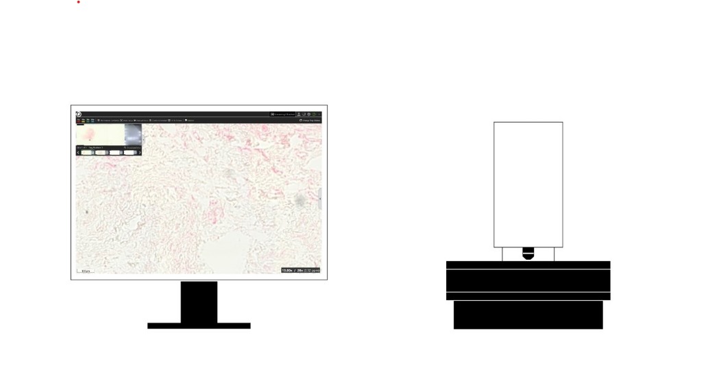
The introduction of digital pathology and remote evaluation platform such Precipoint’s IO:M8 digital microscope and Precicloud storage platform benefits healthcare systems greatly. Even while the upfront costs of buying, setting up, and providing education for the equipment are highly capital-heavy, they cannot be compared to the long-term benefits in terms of health outcomes as well as knowledge transfer. It is possible to completely eliminate the customary, very high cost of expert training overseas, slide, FFPE block/tissue shipping. In addition, the are cost savings as it also eliminates the need for repeat cut sections, and the potential risk of missing a margin of interest leading to delayed or wrong pathology diagnosis. It also facilitates second opinion consultations from partnering pathologists who can access these digital images remotely. Metaphor Laboratory, through its diagnostic value programs is at the forefront of making these solutions accessible and impactful in solving Africa’s healthcare worker attrition burden.
Sequel to this is the utilization of digital images to develop AI algorithms to detect margin of interests, specific biomarkers and/or targets for further therapeutic R&D work. With our central labs in Frederick and Lagos Nigeria, we can support R&D programs ranging from IRB consented biomarker services, Tissue digitization/review services, assay design, development, validation and Genomic services.
References
Griffin, J and Treanor, D. (2017). Digital pathology in clinical use: where are we now and what is holding us back? Histopathology, 70 (1). pp. 134-145. ISSN 0309-0167
Ho J, Aridor O, Parwani AV. (2012). Use of contextual inquiry to understand anatomic pathology workflow: Implications for digital pathology adoption. J Pathol Inform. 3:35.
Nelson, A. M., Milner, D. A., Rebbeck, T. R., Iliyasu, Y. (2016). Oncologic care and pathology resources in africa: survey and recommendations. J Clin Oncol Off J Am Soc Clin Oncol.34:20–6. doi: 10.1200/JCO.2015.61.9767
Randell, R., Ambepitiya, T., Mello-Thoms, C., Ruddle, R.A., Brettle, D., Thomas, R.G., Treanor, D.(2015). Effect of display resolution on time to diagnosis with virtual pathology slides in a systematic search task. J. Digit. Imaging, 28, 68–76.
Stathonikos, N., Nguyen, T.Q., van Diest, P.J. (2020). Rocky road to digital diagnostics: Implementation issues and exhilarating experiences. J. Clin. Pathol..
Wilson, M. L., Fleming, K. A., Kuti, M. A., Looi, L. M., Lago, N., Ru, K.(2018). Access to pathology and laboratory medicine services: a crucial gap. Lancet. 391:1927–1938. doi: 10.1016/S0140-6736(18)30458-6